Back Muscles Anatomy Diagram : 1 - Muscle anatomy diagram printable 12 photos of the muscle anatomy diagram printable muscle anatomy diagram printable, human muscles, muscle anatomy diagram printable.. The deltoid, teres major, teres minor, infraspinatus, supraspinatus (not shown) and subscapularis muscles (not shown) all extend from the scapula to the humerus and act on the shoulder joint. The muscles of the lower back help stabilize, rotate, flex, and extend the spinal column, which is a bony tower of 24 vertebrae that gives the body structure and houses the spinal cord. The extrinsic back muscles, which lie most superficially on the back. Human muscle system, the muscles of the human body that work the skeletal system, that are under voluntary control, and that are concerned with movement, posture, and balance. On this page, you'll learn about each of these muscles, their locations and functional anatomy.
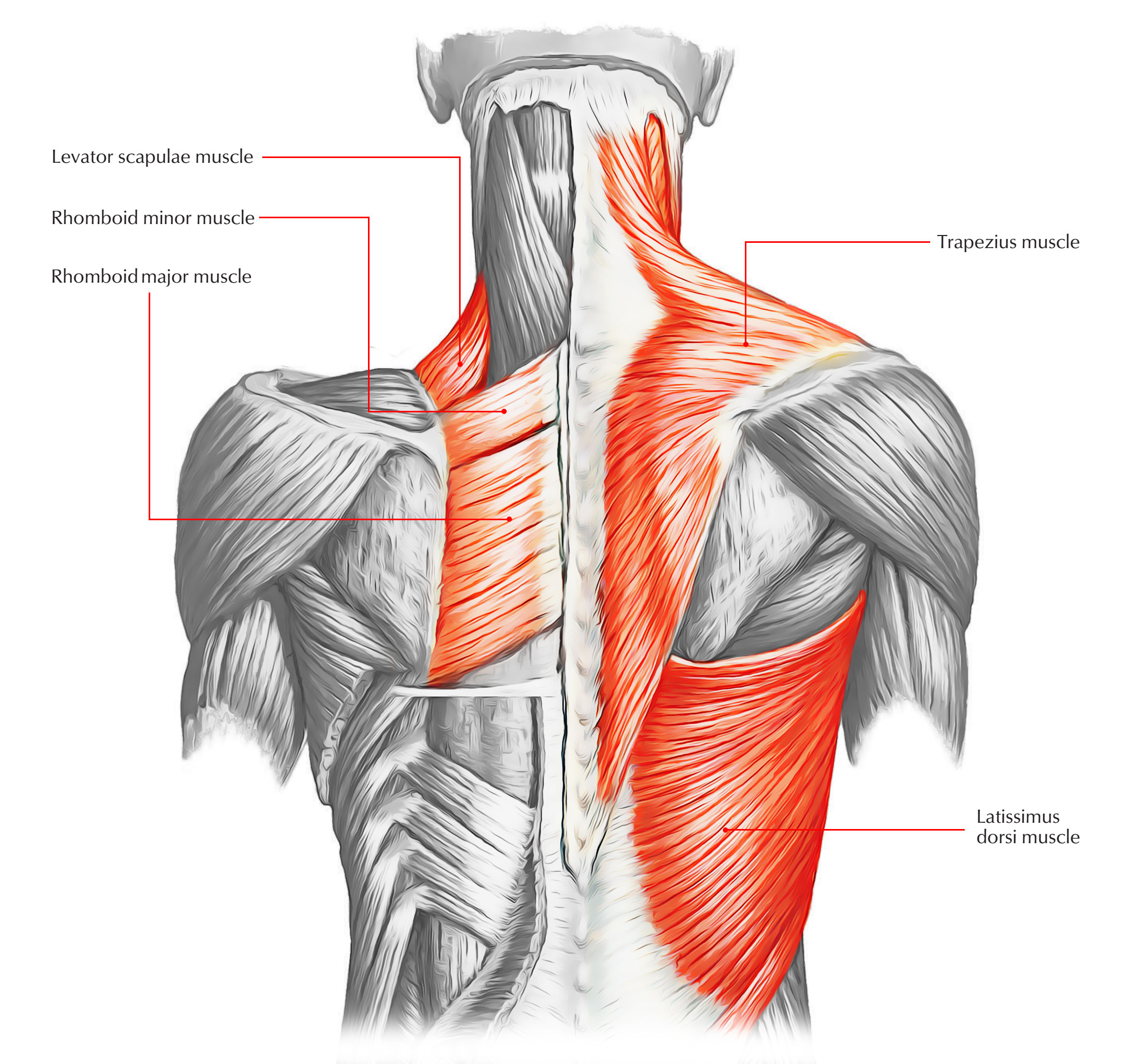
Another common cause of lower back and hip pain is disc injury. The deltoid, teres major, teres minor, infraspinatus, supraspinatus (not shown) and subscapularis muscles (not shown) all extend from the scapula to the humerus and act on the shoulder joint. On this page, you'll learn about each of these muscles, their locations and functional anatomy. The trapezius and latissimus dorsi muscles connect the upper limb to the vertebral column. The back anatomy includes the latissimus dorsi, trapezius, erector spinae, rhomboid, and the teres major.
Back Muscles And Low Back Pain from embed.widencdn.net This human anatomy module is composed of diagrams, illustrations and 3d views of the back, cervical, thoracic and lumbar spinal areas as well as the various vertebrae. Lower back muscle diagram anatomy does degenerative disc disease affect the lower back muscle? Both the deltoid and the trapezius are firmly attached to the spine of the scapula. This image added by admin. You can click the image to magnify if you cannot see clearly. Another common cause of lower back and hip pain is disc injury. They extend and rotate the head and neck. Muscle or ligament strains can occur from repeated use of the muscles, or from improperly or awkwardly lifting heavy objects.
There are three major groups of back muscles:.
The deltoid, teres major, teres minor, infraspinatus, supraspinatus (not shown) and subscapularis muscles (not shown) all extend from the scapula to the humerus and act on the shoulder joint. This human anatomy module is composed of diagrams, illustrations and 3d views of the back, cervical, thoracic and lumbar spinal areas as well as the various vertebrae. Human anatomy for muscle, reproductive, and skeleton. You can click the image to magnify if you cannot see clearly. The bones of the pelvis and lower back work together to support the body's weight, anchor the abdominal and hip muscles, and protect the delicate vital organs of the vertebral and abdominopelvic cavities. Attached to the vertebral column; The quadratus lumborum muscle in the lower back side bends the lumbar spine and aids in the inspiration of air through its stabilizing affects at its insertion at the 12th rib (the last of the floating ribs). Your clients will thank you for it! As you can see, there are also have a spine of scapula deltoid, triceps brachii, latissimus dorsi. This image added by admin. Lumbar aponeurosis and vertebral fascia).—the lumbodorsal fascia is a deep investing membrane which covers the deep muscles of the back of the trunk. Back muscles, like any other muscle in the body, require adequate exercise to maintain strength and tone. The small muscles of the vertebrae (the multifidi and rotators) help rotate, extend, and side bend the back.
While muscles like the gluteals (in the thighs) are used any time we walk or climb a step, deep back muscles and abdominal muscles are usually not actively engaged during everyday activity. Broadly considered, human muscle—like the muscles of all vertebrates—is often divided into striated muscle, smooth muscle, and cardiac muscle. Deep back muscles diagram the superficial layer contains the splenius cervicis and splenius capitis muscles. This is a diagram of the larger and more surface muscles of the low back. Anatomynote.com found anatomy of back muscles diagram from plenty of anatomical pictures on the internet.
Muscles Move And Support The Spine from cloud2.spineuniverse.com The deltoid, teres major, teres minor, infraspinatus, supraspinatus (not shown) and subscapularis muscles (not shown) all extend from the scapula to the humerus and act on the shoulder joint. Broadly considered, human muscle—like the muscles of all vertebrates—is often divided into striated muscle, smooth muscle, and cardiac muscle. On this page, you'll learn about each of these muscles, their locations and functional anatomy. Anatomical diagrams of the spine and back. The muscles of the back are a group of strong, paired muscles that lie on the posterior aspect of the trunk they provide movements of the spine, stability to the trunk, as well as the coordination between the movements of the limbs and the back muscles are divided into two large groups: The extrinsic back muscles, which lie most superficially on the back. Claim your free copy of the client back care guide today. The intricate anatomy of the back provides support for the head and trunk of the body strength in the trunk of the body as well as a great deal of flexibility and movement.
The small muscles of the vertebrae (the multifidi and rotators) help rotate, extend, and side bend the back.
There are three major groups of back muscles:. Both the deltoid and the trapezius are firmly attached to the spine of the scapula. The intermediate layer contains the erector spinae muscles, whose many functions include the extension and lateral flexion of the spine, head and neck. See how exercise helps the back. This is a diagram of the larger and more surface muscles of the low back. Function of the back muscles there are several individual muscles within the back anatomy, and it's important to take a quick look at all of The intricate anatomy of the back provides support for the head and trunk of the body strength in the trunk of the body as well as a great deal of flexibility and movement. The muscles of the back can be arranged into 3 categories based on their location: While muscles like the gluteals (in the thighs) are used any time we walk or climb a step, deep back muscles and abdominal muscles are usually not actively engaged during everyday activity. Link to client back care guide The bones of the pelvis and lower back work together to support the body's weight, anchor the abdominal and hip muscles, and protect the delicate vital organs of the vertebral and abdominopelvic cavities. Female reproductive and digestive system diagram. The superficial back muscles are the muscles found just under the skin.
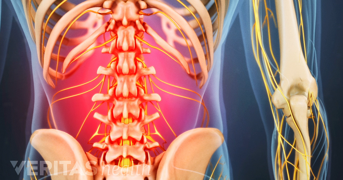
Lumbar aponeurosis and vertebral fascia).—the lumbodorsal fascia is a deep investing membrane which covers the deep muscles of the back of the trunk. A basic understanding of the anatomy of your lower back can help you identify and differentiate a problem that commonly affects this region, such as localized muscle pain or sciatica. Front view of muscles , skeleton , organs , nervous system Human muscle system, the muscles of the human body that work the skeletal system, that are under voluntary control, and that are concerned with movement, posture, and balance. Attached to the shoulder girdle intermediate:
Back Muscles 28 Major Muscles Of The Back Earth S Lab from www.earthslab.com This image added by admin. The intermediate layer contains the erector spinae muscles, whose many functions include the extension and lateral flexion of the spine, head and neck. This is a diagram of the larger and more surface muscles of the low back. Attached to the shoulder girdle intermediate: It contains the osteology, arthrology and myology of the spine and back. The quadratus lumborum muscles (orange, in the image above) are found in the lower back (also called the lumbar area). It is particularly interesting for physiotherapists. The former two groups, superficial and intermediate, are referred to as the extrinsic back muscles.
You can click the image to magnify if you cannot see clearly.
Superficial back muscles, intermediate back muscles and intrinsic back muscles.the intrinsic muscles are named as such because their embryological development begins in the back, oppose to the superficial and intermediate back muscles which develop elsewhere and are therefore classed as extrinsic muscles. It contains the osteology, arthrology and myology of the spine and back. Related posts of muscles of the lower back and buttocks diagram muscle anatomy diagram printable. The deltoid, teres major, teres minor, infraspinatus, supraspinatus (not shown) and subscapularis muscles (not shown) all extend from the scapula to the humerus and act on the shoulder joint. To learn more about the anatomy of the spine, watch this video. Deep muscles of back anatomy biological science picture the muscles of the back can be classified as either deep intermediate and superficial. Attached to the vertebral column; Both the deltoid and the trapezius are firmly attached to the spine of the scapula. On this page, you'll learn about each of these muscles, their locations and functional anatomy. There are three major groups of back muscles:. The latter group is the intrinsic muscle group. The muscles, bones, ligaments, and tendons in the back can all be injured and cause back pain. The intermediate layer contains the erector spinae muscles, whose many functions include the extension and lateral flexion of the spine, head and neck.
Superficial back muscles, intermediate back muscles and intrinsic back musclesthe intrinsic muscles are named as such because their embryological development begins in the back, oppose to the superficial and intermediate back muscles which develop elsewhere and are therefore classed as extrinsic muscles back muscles anatomy. You can click the image to magnify if you cannot see clearly.
Back Muscles Anatomy Diagram : 1 - Muscle anatomy diagram printable 12 photos of the muscle anatomy diagram printable muscle anatomy diagram printable, human muscles, muscle anatomy diagram printable.
Reviewed by MORINX
on
Mei 22, 2021
Rating: 5
buy glibenclamide metformin online acts by decreasing liver glucose production and intestinal glucose uptake. Glibenclamide increases the production of insulin by acting on the pancreas. It decreases the elevated blood sugar level after the meal along with the recommended diet and exercise.


buy glibenclamide metformin online acts by decreasing liver glucose production and intestinal glucose uptake. Glibenclamide increases the production of insulin by acting on the pancreas. It decreases the elevated blood sugar level after the meal along with the recommended diet and exercise.
BalasHapus